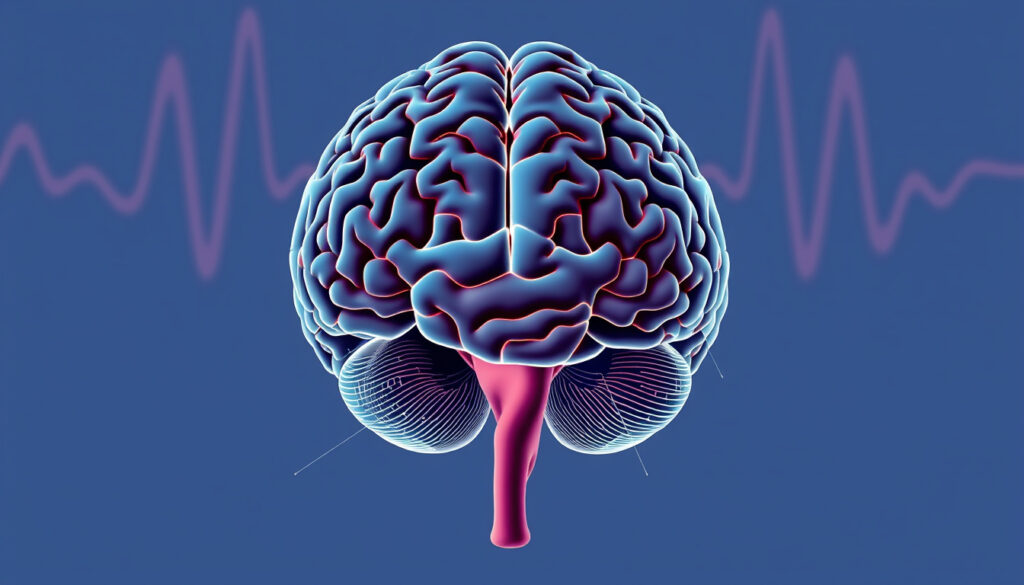
Parts of the Brain: Anatomy, Functions, and Key Structures
Ever wonder why a blow to the back of the head can affect your vision, or why a stroke on the left side impacts the right side of your body? The answers lie in the brain’s intricate architecture, and this guide walks through the major parts from the three main divisions down to the lobes and deeper structures, drawing on the latest from top medical institutions.
Average adult brain weight: 3 pounds (1.4 kg) ·
Number of neurons: 86 billion ·
Percentage of body weight: 2% ·
Oxygen consumption: 20% of total body oxygen ·
Number of lobes in the cerebrum: 4 (frontal, parietal, temporal, occipital) ·
Number of major divisions: 5 (cerebrum, cerebellum, brainstem, pituitary gland, hypothalamus)
Quick snapshot
- The cerebrum is divided into four lobes with distinct functions (Johns Hopkins Medicine (leading academic medical center)).
- The brainstem controls life-sustaining autonomic functions (Mayfield Brain & Spine (neurology specialty clinic)).
- Alzheimer’s disease begins in the hippocampus (Alzheimer’s Society (UK dementia charity)).
- The cerebellum contains more neurons than the cerebrum but is smaller in volume (Johns Hopkins Medicine). (Johns Hopkins Medicine (leading academic medical center))
- The exact mapping of all cognitive functions to specific micro-regions remains an active research area (Queensland Brain Institute, University of Queensland (neuroscience research institute)).
- How consciousness emerges from neural activity is not fully understood. (Queensland Brain Institute, University of Queensland (neuroscience research institute))
- The precise role of the pineal gland in humans beyond melatonin regulation is still debated. (Queensland Brain Institute, University of Queensland (neuroscience research institute))
- The four-lobe model of the cerebrum has remained the standard anatomical framework for over a century, with no fundamental revisions to the lobe boundaries since early 20th-century neuroanatomy (Johns Hopkins Medicine).
- Functional imaging (fMRI) continues to refine sub-region boundaries but does not change the high-level lobe classification.
- Ongoing research using connectomics and high-resolution fMRI will map neural pathways with greater precision, potentially linking specific behaviors to networks spanning multiple lobes (NIDA (National Institute on Drug Abuse, U.S. government research agency)).
- Clinical trials for Alzheimer’s are increasingly targeting early hippocampal changes to slow disease progression. (NIDA (National Institute on Drug Abuse, U.S. government research agency))
Six key measurements, one pattern: the brain is a lightweight organ that demands disproportionate energy resources.
| Fact | Value |
|---|---|
| Brain weight | 3 pounds (1.4 kg) |
| Neurons count | 86 billion |
| Energy consumption | 20% of total body oxygen and calories |
| Number of lobes | 4 |
| Brain parts (major) | 5 |
| Blood supply | Through internal carotid and vertebral arteries |
What are the five major parts of the brain?
Most anatomy textbooks list five major anatomical divisions: the cerebrum, cerebellum, brainstem, pituitary gland, and hypothalamus (Johns Hopkins Medicine). This five‑part model gives a complete top‑to‑bottom view of the brain’s structure.
What is the cerebrum?
- Largest part of the brain — about 85% of its weight (Johns Hopkins Medicine).
- Divided into four lobes: frontal, parietal, temporal, and occipital.
- Controls voluntary movement, senses, language, and reasoning.
Because the cerebrum handles conscious thought and voluntary action, any injury here produces immediately noticeable deficits — a stroke affecting the left frontal lobe can erase a person’s ability to speak, while leaving their understanding of language intact.
What is the cerebellum?
- Located at the back of the brain, below the cerebrum (Johns Hopkins Medicine).
- Coordinates voluntary muscle movements and balance.
- Contains about 50% of the brain’s neurons despite being only 10% of its volume.
What is the brainstem?
- Connects the brain to the spinal cord.
- Composed of the midbrain, pons, and medulla oblongata (Mayfield Brain & Spine).
- Regulates breathing, heart rate, body temperature, and sleep‑wake cycles.
The brainstem is sometimes called the “survival center” because damage to it is often fatal — that’s why a severe blow to the back of the skull can stop a heartbeat in seconds.
What are the pituitary gland and hypothalamus?
- The pituitary gland, a pea‑sized structure at the base of the brain, produces hormones that control growth, metabolism, and reproduction (Johns Hopkins Medicine).
- The hypothalamus sits just above the pituitary and maintains homeostasis (body temperature, hunger, thirst, circadian rhythm).
- Together they link the nervous system to the endocrine system.
The implication: when a patient presents with unexplained weight gain, fatigue, or fertility issues, the first imaging scan often targets this small region — the hypothalamus‑pituitary axis controls nearly every hormonal cascade in the body.
What are the 3 main parts of the brain?
A simpler way to think about brain anatomy is the three‑part model: cerebrum, cerebellum, and brainstem. This is the breakdown most commonly taught in introductory biology (Johns Hopkins Medicine).
What does the cerebrum control?
- Conscious thought, sensation, and voluntary movement.
- Processing of vision, hearing, and touch.
- Higher functions: planning, reasoning, problem‑solving, emotion.
What functions does the cerebellum serve?
- Fine‑tunes motor activity and balance (Johns Hopkins Medicine).
- Coordinates the timing and force of muscle contractions.
- Damage leads to ataxia — uncoordinated movement, not paralysis.
What is the role of the brainstem?
- Manages automatic survival functions: breathing, heart rate, digestion.
- Relays motor and sensory signals between brain and body (Mayfield Brain & Spine).
- Houses the reticular activating system, which regulates arousal and consciousness.
The trade‑off: the three‑part model is easy to remember but omits the endocrine structures (pituitary, hypothalamus) that are functionally critical. That’s why clinicians and neuroanatomists prefer the five‑part division.
What are the 12 parts of the brain?
For a deeper anatomical breakdown, textbooks list 12 key regions that cover the major structures from the cortex down to the brain’s core (Queensland Brain Institute, University of Queensland).
Which structures make up the 12‑part detailed anatomy?
- Frontal lobe, parietal lobe, temporal lobe, occipital lobe, cerebellum, brainstem (midbrain, pons, medulla), basal ganglia, thalamus, hypothalamus, limbic system, pituitary gland, and pineal gland.
- Each has a distinct cellular architecture and set of functions.
What is the corpus callosum?
- A bundle of nerve fibers (white matter) that connects the left and right hemispheres (Mayfield Brain & Spine).
- Enables communication between hemispheres — for example, the left hemisphere’s language center can coordinate with the right’s spatial awareness.
What are the basal ganglia?
- A group of deep brain structures involved in movement control, procedural learning, and habit formation (NIDA).
- Damage here causes movement disorders like Parkinson’s disease (tremor, rigidity, bradykinesia).
What is the limbic system?
- Governs emotion, memory, and motivation.
- Key structures: hippocampus (memory formation), amygdala (fear and emotion), cingulate gyrus (emotional processing) (Queensland Brain Institute).
The limbic system is where emotional trauma gets wired into memory — a car accident survivor’s hippocampus may store the event with an amygdala‑triggered fear response that persists for years, even after the conscious memory fades.
The implication: clinicians must consider multiple brain systems when diagnosing conditions that affect memory and emotion.
“The brain is the most complex part of the human body… The cerebrum is the largest part of the brain and is divided into four sections, called lobes.”
— Johns Hopkins Medicine, Brain Anatomy and How the Brain Works page
What are the 4 main areas of the brain?
The four lobes of the cerebrum — frontal, parietal, temporal, and occipital — form the most clinically useful division of the brain. Each lobe processes a different kind of information (Queensland Brain Institute, University of Queensland).
What is the frontal lobe responsible for?
- Executive functions: planning, reasoning, problem‑solving, emotional regulation.
- Motor planning and voluntary movement (primary motor cortex).
- Personality — a famous 1848 accident (Phineas Gage) showed that destroying the frontal lobe can change a person’s character.
What does the parietal lobe process?
- Sensory information: touch, temperature, pressure, pain (Queensland Brain Institute).
- Spatial awareness and navigation.
- Damage can cause neglect syndrome — a person ignores the opposite side of space.
What functions are linked to the temporal lobe?
- Hearing and language comprehension (Wernicke’s area).
- Memory formation (hippocampus lies within the medial temporal lobe).
- Object recognition and emotional processing (Johns Hopkins Medicine).
What is the occipital lobe’s primary role?
- Vision — receives and processes visual signals from the eyes.
- Located at the back of the head; damage causes blindness or visual hallucinations even when the eyes are healthy (Johns Hopkins Medicine).
The pattern: each lobe processes a single dominant sense (occipital—vision, temporal—hearing), but all lobes work together in networks. A stroke in one lobe rarely produces a pure deficit; patients often show mixed symptoms.
“Your brain contains billions of nerve cells arranged in patterns that coordinate thought, emotion, behavior, movement and sensation.”
— Cleveland Clinic, Brain Anatomy overview
What region of the brain is affected by Alzheimer’s?
Alzheimer’s disease does not attack the brain uniformly — it follows a predictable path, starting in the temporal lobe and spreading outward (Alzheimer’s Society).
Which part of the brain is first impacted?
- The hippocampus, a small seahorse‑shaped structure deep in the temporal lobe, is one of the first areas affected (Alzheimer’s Society).
- The hippocampus is critical for forming new memories — that’s why short‑term memory loss is Alzheimer’s earliest symptom.
How does Alzheimer’s spread through the brain?
- From the hippocampus, the disease moves to the entorhinal cortex, then to the temporal, parietal, and frontal lobes.
- Later stages involve the occipital lobe, affecting vision.
- This spread correlates with the clinical stages: memory loss → disorientation → language and personality changes → loss of motor function.
What are the clinical effects of damage to these regions?
- Hippocampus damage → inability to store new memories.
- Temporal lobe damage → difficulty understanding language (aphasia).
- Frontal lobe damage → poor judgment, apathy, personality changes.
- Parietal lobe damage → spatial disorientation, gets lost easily.
For someone with a family history of Alzheimer’s, early imaging that shows hippocampal shrinkage can be a warning years before memory symptoms appear. The Alzheimer’s Association recommends discussing cognitive screening with a neurologist if you notice persistent short‑term memory lapses.
Early detection of hippocampal changes can significantly improve quality of life through lifestyle interventions and medication.
“The hippocampus is one of the first areas of the brain to be affected by Alzheimer’s disease, leading to problems with short‑term memory.”
— Alzheimer’s Society, How dementia progresses
For a more detailed anatomy of the brain, including diagrams and functional descriptions, you can refer to detailed anatomy of the brain.
Frequently asked questions
What is the purpose of the cerebrospinal fluid?
Cerebrospinal fluid (CSF) cushions the brain and spinal cord, removes waste, and helps regulate intracranial pressure. It is produced in the ventricles of the brain (Johns Hopkins Medicine).
Which part of the brain controls balance?
The cerebellum is primarily responsible for balance and coordination of voluntary movements (Johns Hopkins Medicine).
What happens if the cerebellum is damaged?
Cerebellar damage causes ataxia — a lack of voluntary coordination of muscle movements, including gait, speech, and eye movements. Patients may have trouble walking or reaching for objects (Mayfield Brain & Spine).
How do the left and right hemispheres differ in function?
The left hemisphere is typically dominant for language and logic, while the right hemisphere excels at spatial awareness and emotional processing. They communicate through the corpus callosum (Mayfield Brain & Spine).
What part of the brain regulates breathing?
The brainstem — specifically the medulla oblongata and pons — controls the rhythm and depth of breathing (Mayfield Brain & Spine).
What is the corpus callosum and what does it do?
The corpus callosum is a thick band of nerve fibers that connects the left and right hemispheres, allowing them to communicate and coordinate responses (Mayfield Brain & Spine).
Which region of the brain is involved in fear and emotion?
The amygdala, part of the limbic system deep in the temporal lobe, plays a key role in processing fear, anxiety, and emotional memory (Queensland Brain Institute).
What is the role of the thalamus?
The thalamus acts as a relay station, routing sensory signals (except smell) from the body to the appropriate areas of the cerebral cortex (Johns Hopkins Medicine).
For more on related health topics, see Low Platelet Count Meaning: Thrombocytopenia Explained and What Is the Best Liver Food? Top Foods for Repair & Detox.
For anyone caring for a loved one with Alzheimer’s, the decision to seek early intervention is clear: talk to a neurologist about cognitive testing, or watch daily memory lapses slowly erode independence.
More related posts
 Google Image Search Reverse – How to Use on Desktop and Mobile
 Becky Lynch: Real Name, Baby Daddy, Irish Roots & Happy Gilmore
 Breaking Bad: Why It’s So Popular, Characters & More
 Kim Woo Bin: Relationship, Cancer Diagnosis, and Marriage Update
 Korean Won to CAD – Live Rate History and Converter Guide
 Natalie Viscuso: Age, Job, Baby with Henry Cavill (2025)
 Who Won Dancing with the Stars 2024? Winners for US and Ireland
 Kylie Minogue: Biography, Partner, Face Surgery, Cancer & More